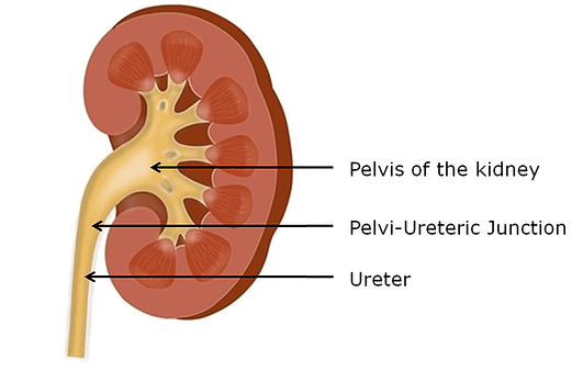
Pelvi-Ureteric Junction (PUJ) Obstruction
PUJ is the junction between the pelvis of the kidney and the ureter (the tube between the kidney and the bladder).
Symptoms of PUJ obstruction
PUJ obstruction refers to a narrowing of the junction, resulting in obstruction to the flow of the urine from the kidney to the ureter.
The condition affects approximately one person in every 1000 adults and tends to occur more in men.
Occasionally PUJ obstruction causes no symptoms or problems and is only discovered by chance when the patient is having a scan for another condition. Alternatively it can cause:
- Recurrent episodes of loin pain which tends to worsen after drinking especially alcohol.
- Kidney infection (pyelonephritis).
- Kidney stones.
- Lump or swelling in the kidney area.
- Damage to the kidney as a result of either high pressure in the renal pelvis, kidney infection or formation of kidney stones.

The condition affects approximately one person in every 1000 adults, and tends to occur more in men.
Causes of PUJ obstruction
Treatments for PUJ obstruction
Most commonly PUJ obstruction is caused by a congenital weakness in the wall of the junction. Sometimes there is an extra blood vessel pressing on the junction from outside causing or contributing to the obstruction (known as accessory vessel).
Occasionally PUJ obstruction is acquired as a result of either formation of a scar tissue following passage of a kidney stone or very rarely due to cancer.
Diagnosis of PUJ obstruction
This includes blood tests, urine test and scans. CT scan is commonly used to assess the anatomy and the structure of the renal pelvis and a special nuclear medicine scan called MAG3 scan is used to confirm the obstruction and also to assess the function of the kidney.
If there is severe kidney infection as a result of the obstruction, then the kidney must be drained as a matter of urgency with insertion of a temporary ureteric stent or nephrostomy tube before any definitive treatment.
There are several treatment options for PUJ obstruction and these will be discussed with you; these include:
Active surveillance with careful observation with repeated scans.
Surgery
- Laparoscopic (keyhole) pyeloplasty.
- Endopyelotomy (with laser).
- Long-term ureteric stent.
- Laparoscopic (keyhole) nephrectomy may be considered if the kidney is not functioning.


